When Choosing the Best Medical Education Models, Materials Matter
The first generation of bone models made their appearance in the mid-1970s, and they began an evolution that has led to modern medical education model materials that are better than ever. The first iteration of these models lacked critical detail such as the medullary cavity. The largely fiberglass models offered little in the way of hands-on functionality or realism, instead focusing on the fundamentals. Since then, the materials used in medical education models have transformed to not only replicate human skin, bone, muscle, and tissue, but they also have dynamic functions that make them essentials in modern instruction across specialties and procedures.
Adding Function to Form
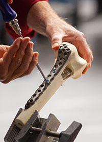
The addition of essential features has meant that medical education models can now be used for advanced instruction, such as inserting screws, administering injections, and treating fractures. The journey that began with those early 90s models has since led to engineered education solutions superior to cadavers.
Today’s models are cheaper—human femurs, tibia, and humeri cost around four times as much as an advanced model—and they are more robust than cadavers. They can be designed to replicate any presentation or procedure, raise no ethical concerns, and don’t skew toward elderly specimens, unlike human samples.
The direct descendants of the initial class of models have also evolved dynamic features beyond the materials used in their composition, such as haptic feedback and progressive buildability, that make them potentially more useful in instruction than some of the DIY 3D printable resources currently being explored. There is a clear potential use for such highly customizable models in personalized surgical preparation, but until the base materials can better replicate the human reality, those 3D printed models may be less practical than engineered solutions.
There are several examples of advanced medical education materials models that clearly demonstrate the importance of getting a realistic performance and response from training resources.
Composite Bone – Varus Deformity
Human bones are not composed of uniform material. The six types of bone vary in density, internal structure, and the composition of fibrous and cellular layers, and this determines the appropriate intervention for treatments across the body. The most common surgical treatment of “varus knee,” for example, is a high tibial osteotomy. A model that is used to train students on the procedure needs to realistically recreate the performance, anatomy, and tensile strength of the tibia so that it can withstand the cutting and reshaping of the bone needed for a successful outcome.
Such advanced models can also effectively recreate the load and motion properties of bone to give students hands-on experience in non-surgical fields such as osteopathy and chiropractic.
Lifelike Skin and Tissue – Suture Training Models
Quality educational training model materials make it easier to transfer core skills to the operating room. Suturing is one such critical skill that can impact the effectiveness of a wide range of surgical interventions but requires a specific set of resources to master. The best models have highly accurate skin layers, blood vessels, neurological networks, follicles, fat, and muscle tissue that closely resemble human anatomy.
If your model does not conform to the realities students will encounter in the operating room, then you’re not preparing them to efficiently navigate the basics such as continuous and interrupted suture patterns, two-level closure, or critical intradermal sutures that can define patient outcomes.
Radiopaque Materials – Fluoroscopy Models
The best medical education model materials serve practical ends. Such is the expertise and range of modern model engineers that they can use these advanced materials in collaboration with medical instructors to produce resources specific to any procedure. Fluoroscopy models, for example, capitalize on modern radiopaque materials to recreate the live imaging used in angiograms, barium x-rays, orthopaedic surgery, and the insertion of devices such as stents.
Radiopaque materials that block x-rays can be used to create models of any part of the skeleton, making them powerful training tools for any imaging application.
These dynamic materials are a dramatic improvement on the fiberglass models that came to prominence 30 years ago. They can be applied to a range of models to aid medical education across any medical field.
The Right Materials for Your Field
The advanced training model materials listed above, and many more, can be applied to as many scenarios as there are best practice procedures. They can be incorporated into models of any part of the body and used in combination with dynamic features to produce models capable of recreating complex skills and techniques.
More importantly, these materials can be applied to models for any field:
- Chiropractic: Realistic models better recreate the response of bone, vertebrae, and muscle to spinal column misalignment corrections. The best models will also mimic the range of movement through the lumbar region.
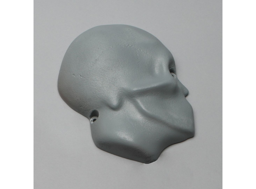
- Neurosurgery: Authentic composite bone materials can be added to both open and closed models, helping students master the tools and techniques necessary for the most common craniotomy procedures.
- Orthopaedic: It is important that model materials authentically reproduce the movement of joints and the difficulties of navigating them with arthroscopic devices.
- Pediatric: Childhood models have a unique burden of realism, as juvenile bone performs differently than adult versions most encountered in cadavers. From scoliosis to greenstick fractures, these models require special attention in their construction.
- Rheumatology: The huge variety of diseases and conditions treated by rheumatologists means they require training models that suit a range of methods. From radiopaque x-ray simulations to arthroscopic models that hone psychomotor skills, the best models prepare trainees for real environments.
These advanced materials are the result of more than 30 years of medical education model evolution. They represent the best resources medical instructors have ever had at their disposal.
Make the Most of Medical Education Models Materials
Unlike your educational predecessors of previous decades, your ambitions as an instructor are not determined by the quality of resources at your disposal. There are authentic, functional, and fully customizable models to suit any best practice instruction.
By partnering with the right engineering team, you can create models that not only replicate the human body, but also the pressures, intricacies, and challenges of providing best possible outcomes to patients. Today’s models are effective enough to make any skill teachable and transferable.
Sawbones engineers have been at the cutting edge of educational model materials development, helping create the broadest range of products instructors have ever had at their disposal. View our catalog, or get in contact with us online or by phone at 206-463-5551 to learn more.

If you're seeking something you can't find on our website, our sales team is happy to help. We can either direct you to the right model or provide a free quote on the right custom project to meet your needs. Discover options with our clear bone models, laminated blocks, custom displays, or other machining projects.

