
5 Femur Bone Model Features to Best Show and Explain Fracture Patterns
Femur fractures are serious injuries afflicting 10-21 per 100,000 people per year. These are almost always a result of a high-energy mechanism injury, like a motor vehicle accident, so it's something that trauma surgeons and emergency room workers will often contend with. On top of that, these fractures require extensive follow-up, so many other medical professionals like family practitioners, physical therapists, and more will need a thorough understanding of them. Training using specific femur bone model features is necessary for a wide range of specialties.
Ideally, bone models will help individuals understand the specific type of injuries common in this area. Many will require extensive surgery and medical intervention to resolve. Suitable bone models for medical training will help prepare trainees for a wide range of issues they'll run into when dealing with this part of the body.
Common Femoral Shaft Fracture Patterns
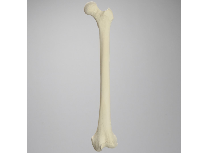
The femur is the longest and strongest bone in the human body. It has to be for the amount of weight and stress it supports. However, that strength doesn't guarantee protection. When injuries occur, they're often severe. Four types of fractures are common:
Comminuted |
Open |
Oblique |
Transverse |
|
The comminuted fracture is extremely serious, as it causes the bone to break into three or more pieces. Surgery is almost always required—sometimes in multiple sessions to remove the bone fragments and insert hardware for stability. |
Open fractures are complex, as they also involve soft tissue damage. In these, the fracture punctures the skin. The patient is at extreme risk of infections and complications during healing. Open fractures frequently accompany comminuted ones. |
This fracture is curved or at an angle—which can complicate healing. It's also not unusual to see fragments due to the abnormal shape of the break. Surgery may be indicated to remove loose pieces and set the bone in place. |
The transverse fracture occurs straight across the femoral shaft, putting stability at risk. The most common procedure for resolving one involves using hardware to set the bone in place. |
The handling of a femoral fracture is almost always an extended process, resulting in permanent impairment. Even in instances where residents won't treat the injury itself, they may have to manage ongoing effects from it in their everyday work with patients.
5 Femur Bone Model Features for Fracture Practice
Femurs are an easy bone to identify and are often used in early medical education because the size and shape make them suitable for demonstration purposes. However, when it comes to learning how to treat a femur injury—and especially a fracture—it's wise to consider more advanced medical education model options. Femur bone model features to consider include:
1. Radiopaque properties
Radiopaque properties are necessary for teaching some types of imaging, as they block x-rays and keep them from passing right through the material. A standard bone model placed in an x-ray machine wouldn't be a useful teaching tool, as it doesn't have the same radiopaque properties as bone. Follow-up imaging is a necessity in femur fractures in order for the practitioner to ensure the bone is healing properly. Radiopaque bone models allow students to learn imaging techniques and identify fracture patterns.
2. Comorbidities
While fractures occur in healthy bones, this will not be the case every time. Practitioners need a visual of common comorbidities that occur outside the injury, like arthritis or osteoporosis. These conditions complicate recovery and will require consideration within the overall plan of care.
3. Realistic cancellous bone
The internal tissue within the bone is far different from its surface.Trainees need to understand this spongy material, how it's impacted in an injury, and its overall effect on recovery. It's also different to work with when compared to the compact bone surrounding it, so a realistic texture for practicing setting and hardware insertion is vital.
4. Realistic compact bone
Compact hard bone is unique and requires special handling when setting fractures and drilling holes. A realistic cortical shell model can recreate the feel of compact bone while allowing for easier drilling and handling for residents.
5. Transparent features
Early in medical training, it's helpful to have fracture models with transparency so students can see within the bone without the need to cut into it. They can view the inner cancellous material, and see how certain conditions and comorbidities affect the internal structure. This is especially helpful in intramedullary training.
Some femur bone model features are more valuable than others when instructors attempt to teach the treatment of common fractures. These are serious injuries with long recovery times, so it's not just surgeons who will need to understand them. Training for palliative long-term care is also essential. Advanced features of femoral models will help these trainees gain the knowledge they need to improve patient outcomes, regardless of their chosen specialty.
Sawbones offers the best bone model features for detailed femur fracture training. For more information on our offerings or to talk about custom training models, contact us at 206-463-5551.

If you're seeking something you can't find on our website, our sales team is happy to help. We can either direct you to the right model or provide a free quote on the right custom project to meet your needs. Discover options with our clear bone models, laminated blocks, custom displays, or other machining projects.

Work with the industry leader in medical training products!
For four decades, Sawbones has offered the best medical training products for doctors, educators, sales specialists and more. Contact us to learn more about our high-quality models!
1 (206) 463-5551Contact Us!


