How Your Elbow Moves: Explaining Flexion, Extension, and More
Introduction
Understanding how your elbow moves is crucial, whether you're a medical professional or a student. At Sawbones, we offer top-notch anatomical models that make the complex mechanics of the elbow easier to understand and teach. Whether you're a surgeon, a medical student, or involved in developing medical devices, our models can enhance your practice and knowledge. This article explores the intricate movements of the elbow, focusing on flexion, extension, and the muscles involved, providing valuable insights for various audiences, including medical professionals, educators, and students.
Anatomy of the Elbow
The elbow is a sophisticated hinge joint that connects the upper arm bone (humerus) to the forearm bones (radius and ulna). It's essential for many daily activities and intricate medical procedures. Using Sawbones anatomical models, you can visually explore these components:
- Humerus: The upper arm bone forming the upper part of the elbow joint.
- Radius: The forearm bone on the thumb side, crucial for rotational movements.
- Ulna: The forearm bone on the pinky side, forming the hinge of the elbow.
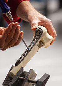
Our models allow for hands-on learning, showing how these bones interact realistically.
Types of Movements
Understanding the types of movements your elbow can perform is essential for diagnosing and treating various conditions. Elbow Models by Sawbones accurately depict these movements:
- Flexion: This movement decreases the angle between the forearm and the upper arm, facilitated by the biceps brachii muscle. Flexion allows you to bring your hand toward your shoulder.
- Extension: This movement increases the angle between the forearm and the upper arm, straightening the arm. The triceps brachii muscle is the primary facilitator of this action.
With our models, you can observe these movements in real-time, making it easier to understand the mechanics involved.
Muscles Involved
The muscles around the elbow are crucial for its movement and stability. Here are the primary muscles involved, which can be studied using Sawbones models:
- Biceps Brachii: Located at the front of the upper arm, responsible for flexion and supination of the forearm.
- Triceps Brachii: Located at the back of the upper arm, responsible for extension of the elbow.
- Brachialis: A muscle beneath the biceps that assists in elbow flexion.
- Brachioradialis: A forearm muscle that contributes to flexion, especially when the forearm is in a mid-pronated position.
Our models provide detailed representations of these muscles, allowing for a thorough examination of their roles in elbow movements.
Common Injuries and Prevention
Elbow injuries are common, particularly among athletes and individuals engaged in repetitive arm movements. Common injuries include:
- Tennis Elbow (Lateral Epicondylitis): An overuse injury causing pain on the outer part of the elbow.
- Golfer’s Elbow (Medial Epicondylitis): Similar to tennis elbow but affects the inner part of the elbow.
- Elbow Fractures: Breaks in the bones of the elbow due to trauma or falls.
To know more about elbow fractures, check out our blog. - Dislocations: When the bones of the elbow are forced out of alignment.
Using Sawbones models, medical professionals can better understand these injuries, how they occur, and the best methods for treatment and prevention.
Prevention Tips:
- Proper Warm-Up: Always warm up before engaging in physical activities.
- Strengthening Exercises: Regular exercises to strengthen the muscles around the elbow.
- Ergonomic Tools: Use tools and equipment that reduce strain on the elbow during repetitive tasks.
Final Words
For medical professionals, educators, and students, understanding the detailed mechanics of the elbow is vital. Sawbones anatomical models offer a realistic and practical way to study and teach these concepts. By comprehending the anatomy, types of movements, and the muscles involved, you can better diagnose, treat, and educate others about elbow-related issues. This knowledge not only enhances clinical practice but also contributes to effective teaching and learning in medical education.

